Center for Cancer Research Featured News Archive
View an archive of research highlights and featured publications from the Center for Cancer Research.
2021
Cancer Center Researchers Included on Highly Cited Researchers 2021 List
We are proud to share that 28 Mass General Cancer Center researchers have been named to the Highly Cited Researchers list of 2021 by the Institute for Scientific Information at Clarivate.
This list identifies and celebrates exceptional individual researchers who are having a significant impact on the research community as evidenced by the rate at which their work is being cited by their peers. The research they have contributed is fueling the innovation, sustainability, health and security that is key for the future. Read more.
Fibroblasts could serve as new key to enhancing personalized treatment for lung cancer patients
Three subtypes of cancer-associated fibroblasts (CAF) could guide the design of personalized treatment for lung cancer patients, according to a new study in Cancer Cell led by researchers at Massachusetts General Hospital.
“We need a new approach to characterize CAFs. Importantly, we need to gain a comprehensive understanding of different CAFs’ biological functions and their clinical significance,” says lead author Haichuan Hu, MD, PhD of the Hata lab at the Center for Cancer Research. View the press release.
Researchers identify mechanisms of resistance to drug for triple-negative breast cancer
Massachusetts General Hospital researchers, including Aditya Bardia, MD, MPH and Leif Ellisen, MD, PhD (Ellisen lab), have identified for the first time how a highly aggressive form of breast cancer can evade one of the most powerful and effective drugs used to treat it, reporting their findings in Cancer Discovery, a journal of the American Association for Cancer Research. The findings could help improve therapy and ultimately prolong survival for patients with metastatic triple-negative breast cancer. View the press release.
Researchers find immune cell “hubs” in certain tumors
Immune cells in some human colorectal tumors congregate in clusters, scientists report in Cell. The researchers, from MGH, the Broad Institute of MIT and Harvard, MIT, the Evergrande Center for Immunologic Diseases at Brigham and Women’s Hospital and Harvard Medical School, and Dana-Farber Cancer Institute say that these “hubs” might be likelier to respond to immunotherapies.
Nir Hacohen, PhD, who is the David P. Ryan, MD, Endowed Chair in Cancer Research at MGH and director of the Center for Cancer Immunology, is co-senior author of the study. View the press release.
Researchers pinpoint how PARP inhibitors combat BRCA1 and BRCA2 tumor cells
In a study published in Genes and Development, a team of Mass General researchers describe how an important class of anti-cancer drugs called PARP inhibitors works, a finding that could help improve treatment and prolong survival for patients with breast cancer and other malignancies.
As Zou Lee, PhD, scientific co-director of the Mass General Cancer Center, and colleagues found, PARP inhibitors work by creating gaps in tumor-cell DNA that remain present through multiple cell cycles (the process by which cells replicate: grow, divide, repeat). They also found that BRCA1/2 mutant cancer cells cannot respond to these gaps and therefore fail to repair properly, leading to the death of tumor cells. View the press release.
The mutation-independent immunogenic effect of environmental carcinogens blocks cancer metastasis

Emerging evidence indicates that immunogenicity (i.e., improved response to immunotherapies) is a hallmark of cancers caused by exposure to environmental carcinogens. But in the past, science has focused mainly on the mutations caused by these exposures in a patient’s DNA as the reason for the immune attack. In a June 2021 paper published in Science Advances, Li et al. describe a distinct consequence of exposure to carcinogen that can have significant immunologic implications: the nongenetic induction of a chemokine called CCL21 in breast cancer cells that developed following an exposure to a chemical carcinogen. This finding could open the door to therapeutic interventions to improve the response of nonimmunogenic “cold” tumors to current cancer immunotherapies. View the press release.
More news from the Demehri lab:
- Common moles could serve as players in battling melanoma and preventing its recurrence
- New Insights on How Inflammatory Molecule Contributes to Skin and Pancreatic Cancers
Mass General Cancer Center to Host Inaugural Arthur and Sandra Irving Cancer Immunology Symposium, March 22-24
The Mass General Cancer Center will host the Arthur and Sandra Irving Cancer Immunology Symposium from March 22-24, 2021, virtually bringing together 14 accomplished faculty mentors who will discuss their research, discoveries, and distinguished careers in cancer immunology with 44 talented young scientists and physicians from around the world. Learn more.
Cosmetic laser may boost effectiveness of certain anti-cancer therapies
Use of a cosmetic laser invented at Massachusetts General Hospital may improve the effectiveness of certain anti-tumor therapies and extend their use to more diverse forms of cancer. The strategy was tested and validated in mice, as described in a study published in Science Translational Medicine.
In an attempt to expand the benefits of immune checkpoint inhibitors for additional patients, a team led by CCR faculty member David E. Fisher, MD, PhD, director of the Mass General Cancer Center’s Melanoma Program and director of MGH’s Cutaneous Biology Research Center, conducted experiments in mice with a poorly immunogenic melanoma that is not hindered by immune checkpoint inhibitors. The researchers found that exposing the melanoma cells to ultraviolet radiation caused them to take on more mutations, which made immune checkpoint inhibitors more effective at boosting the immune response against the melanomas. View the press release.
More news from the Fisher lab:
2020
Study uncovers two phases of infection in patients with severe COVID-19 pneumonia
New research led by investigators at Massachusetts General Hospital and published in Nature Communications provides insights that could help improve treatment strategies for infected patients.
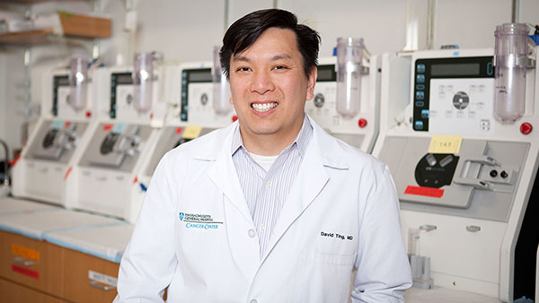
To analyze SARS-CoV-2 at the tissue level, the scientists examined autopsied material from 24 patients who succumbed to COVID-19. “We used a method called RNA in situ hybridization to visualize the actual SARS-CoV-2 virus in human lung specimens. This assay is now a clinical test being used at MGH to understand what tissues can be infected by the virus,” explains co-author David T. Ting, MD, associate clinical director for Innovation at the Mass General Cancer Center and an assistant professor of Medicine at Harvard Medical School. View the press release.
CCR Investigators Named Bits to Bytes Program Awardees
Congratulations to CCR faculty Raul Mostoslavsky, MD, PhD, Nabeel Bardeesy, PhD, Gad Getz, PhD, and Leif Ellisen, MD, PhD, who along with Keith Flaherty, MD have been awarded Bits to Bytes program funding from the Massachusetts Life Sciences Center (MLSC) for their project "An Integrative Platform to Understand and Exploit Cancer Metabolism."
MLSC launched Bits to Bytes in 2019 to provide grants for projects that generate and analyze large datasets to answer pressing life science questions, and to attract and train data scientists in the Commonwealth. View the press release.
Lee Zou, PhD and Raul Mostoslavsky, MD, PhD Named Scientific co-Directors
Congratulations to Lee Zou, PhD and Raul Mostoslavsky, MD, PhD who will serve as Scientific co-Directors of the Mass General Cancer Center and Center for Cancer Research.

After nine years as the Scientific Director of the Mass General Cancer Center and Center for Cancer Research, Dr. Nick Dyson is stepping down from his role. Succeeding him are Lee Zou, PhD and Raul Mostoslavsky, MD, PhD, who will serve as Scientific co-Directors. Both of these highly accomplished researchers are ready to ensure that Mass General Cancer Center continues on its outstanding trajectory of discovery and innovation in cancer research. Read more.
Ultrahigh-throughput magnetic sorting of large blood volumes for epitope-agnostic isolation of circulating tumor cells
A team at Massachusetts General Hospital has devised a new ultrahigh-throughput microfluidic chip (the LPCTC-iCHIP) that improves sensitivity of CTC-based assays 100-fold as compared to the existing approaches. The team’s paper appeared recently in the May 2020 issue of Proceedings of the National Academy of Sciences.

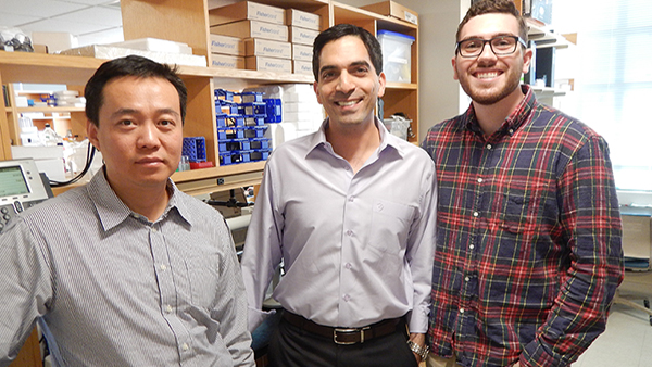
“Drs. Mehmet Toner, Daniel Haber, and Shyamala Maheswaran applied the precision of microfluidics to rare cell sorting and helped establish the field of liquid biopsy research about ten years ago,” says Avanish Mishra, PhD, a research fellow at the Mass General Cancer Center and Harvard Medical School, who was the co-lead author on the LPCTC-iCHIP project along with Taronish Dubash, PhD. “With this novel new technology and our unique process using the leukopak, we transform CTC screening from a 10 mL blood sample to whole human blood; hoping to bring highly reliable liquid biopsies into the clinic.” View the press release.
Genomic characterization of human brain metastases identifies drivers of metastatic lung adenocarcinoma
Cancer researchers at Massachusetts General Hospital and affiliated institutions have identified changes in lung cancer-promoting genes that may allow the disease to metastasize to the brain. Their work, which points to possible therapies for preventing or treating brain metastases from lung cancer, is described in the March 2020 issue of Nature Genetics.

“It’s a terrible complication of lung cancer, because it’s associated with significant mortality, and the median survival in this patient population is on the order of months. Unfortunately, the treatment options are limited and there is no established standard of care for most patients with brain metastases from lung adenocarcinoma,” said principal investigator Priscilla K. Brastianos, MD, from the Mass General Cancer Center. View the press release.
Deregulation of ribosomal protein expression and translation promotes breast cancer metastasis
Hormone receptor-positive breast cancer can spread throughout the body via the bloodstream as circulating tumor cells, or CTCs, which eventually reach distal (remote) body sites to form metastatic tumors. An increase in ribosomes, the protein-making machinery found in every living cell, increases their potential to form metastasis, report investigators from Massachusetts General Hospital Cancer Center and Harvard Medical School. The findings were published in a March 2020 issue of Science.
“The Haber/Maheswaran lab has been one of the pioneers in being able to isolate, analyze and now use CTCs that are derived from patient blood samples. They offer many advantages and opportunities for understanding the metastatic pathways that are involved in advanced breast cancer,” says investigator Douglas S. Micalizzi, MD, PhD, from the MGH Cancer Center. View the press release.
Tumor sequencing has traditionally focused on protein-coding genes, yet these only comprise roughly one percent of the human genome. The remainder contains important regulatory sequences that tightly control gene expression and production of proteins, making sure genes are turned on at the right time in the correct cells.

The ICGC/TCGA Pan-Cancer Analysis of Whole Genomes Consortium, a joint effort of over 1,300 researchers from 37 countries, published a comprehensive in-depth analysis of more than 2,600 primary tumor genomes in the Feb 2020 issue of Nature. As part of this effort, CCR faculty members Gad Getz and Esther Rheinbay, together with their colleagues, studied the landscape of protein-coding and non-coding regulatory driver events in cancer. Their study presents a novel strategy to analyze recurrent driver events in whole cancer genomes, and shows that although there are clear drivers in non-coding regions, the majority of driver genetic changes alter proteins. This work also highlights that there are still challenges in finding genetic drivers of cancer and that we will need to sequence many more tumors to fully map and understand the genetic causes underlying each individual patient’s disease. View the press release.
TATR protects the genome by sensing R loops

R loops are transcription intermediates that contain DNA:RNA hybrids. In cancer cells, R loops often accumulate at aberrantly high levels, leading to genomic instability and DNA damage. In a February 2020 paper published in Molecular Cell, Matos and colleagues in the Zou laboratory reported that the ATR kinase is a key sensor of R loops and a suppressor of R loop-associated DNA damage. This paper reveals the molecular mechanisms by which human cells, especially cancer cells, use ATR to cope with R loops in their genomes, suggesting that inhibition of ATR may be an effective strategy to selectively kill cancer cells harboring high levels of R loops.
2019
Immunity to Commensal Virome Blocks Cancer
In an October 2019 paper published in Nature, Strickley, Messerschmidt et al. investigated the role commensal human papillomaviruses (HPVs) play in the development of skin cancer. Using a novel skin colonization model, they show that adaptive immunity against papillomavirus infection is able to protect the host from developing cancer. In stark contrast to the established “hit and run hypothesis,” which postulates that skin HPVs initiate cancer and are later lost, this work elucidates the significant and previously unrecognized beneficial contributions of skin-resident HPVs. Considering the >100-fold increase in skin cancer risk among immunosuppressed patients, these findings potentiate novel therapeutic interventions to prevent the development of skin cancer in this high-risk population. Furthermore, these results establish a new field of investigation into the beneficial contributions of viruses that live in our skin and other organs. View the press release.
CAR-T cells secreting BiTEs circumvent antigen escape without detectable toxicity.

Building on their research showing that an exciting new form of immunotherapy for cancer has activity in patients with glioblastoma, the most common and most deadly form of brain cancer, Mass General investigators have created a new method that could make immune therapy more effective again brain tumors and expand its use against other types of solid tumors. Their study is published in the July 2019 issue of Nature Biotechnology. View the press release.
An Integrative Model of Cellular States, Plasticity, and Genetics for Glioblastoma.

The researchers profiled gene expression in more than 24,000 tumor cells from 20 adult and eight pediatric glioblastoma patients and also analyzed glioblastoma models in the lab. They found four glioblastoma cellular states, which each has a unique gene expression program and together help account for the large variation in the disease. The scientists then used the single-cell data to reanalyze glioblastoma data from The Cancer Genome Atlas, and identified genetic alterations associated with each of the four states. The results, published in Cell, also show that the cells are remarkably flexible or plastic — that is, they can switch between the four states. This shape-shifting ability could help explain why these cancer cells are so difficult to kill with drugs and help inform the development of better therapies for glioblastoma. The research was led by co-first authors Cyril Neftel, Department of Pathology at Massachusetts General Hospital (MGH) and Broad Institute; Julie Laffy, Weizmann Institute of Science; Mariella Filbin, Department of Pathology at MGH, Broad Institute, and Dana-Farber Institute; Toshiro Hara, Salk Institute for Biological Studies; and co-senior authors Itay Tirosh, Weizmann Institute of Science, and Mario Suvà, Department of Pathology at MGH and Broad Institute. View the press release.
Visualizing Engrafted Human Cancer and Therapy Responses in Immunodeficient Zebrafish.
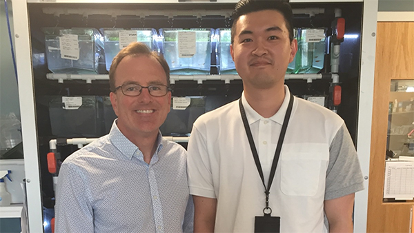
In a June 2019 paper published in Cell, Yan, Langenau and coworkers show that they can engraft a wide array of human tumors in optically clear zebrafish. The advantages of this system for imaging mean that it is readily possible to visualize dynamic changes in single engrafted cells. The authors have used their zebrafish models to show the preclinical efficacy of a drug combination of the PARP inhibitor Olaparib and the DNA-damaging agent Temozolomide for rhabdomyosarcoma. The ability to grow patient-derived tumors in zebrafish will likely open many new avenues for personalized medicine.
Passenger hotspot mutations in cancer driven by APOBEC3A and mesoscale genomic features.
The DNA of cancer cells contains many mutations. A few of these mutations are called "driver mutations" because they were responsible for changing the behavior of the cells to make them multiply out of control and form a tumor. But the vast majority of mutations in the cell had no effect and are just "along for the ride" - these are called "passenger mutations". Telling driver mutations apart from the far more numerous passenger mutations can be very challenging. One commonly used approach is to look for exactly the same mutation occurring in many different patients' cancers. This "recurrence" approach has been very successful over the past decade at identifying cancer driver genes and mutations. However, it assumes that every recurrent mutation ("mutation hotspot") is a driver hotspot. In this 2019 paper published in Science, MGH researchers Dr. Lee Zou and Dr. Michael Lawrence took a new critical look at this assumption. Combining the power of biochemistry and bioinformatics, they and their co-authors showed that some hotspots are actually "passenger hotspots," a term that would be seen as contradictory given the current thinking of the field. These passenger hotspots result from the very specific preferences of APOBEC3A, a DNA-mutating enzyme that is often upregulated in cancer cells. They showed that APOBEC3A has a very strong preference for mutating C nucleotides exposed in a short loop at the end of a DNA "hairpin". DNA hairpins form when nearby palindromic DNA sequences in one DNA strand pair to each other instead of their complementary partner on the opposite DNA strand. This generates a perfect substrate for APOBEC3A to attack, and leads to the formation of passenger hotspots, which are recurrently mutated in many patients' cancers, despite contributing nothing to the development or fitness of the cancers. These results show that some previously nominated cancer drivers (e.g. a hotspot in the gene MB21D2) are actually passenger hotspots, and would likely not be worthwhile targets for further study. Incorporating these insights into driver discovery algorithms will improve our ability to detect the true drivers of cancer.
Transcriptome-wide off-target RNA editing induced by CRISPR-guided DNA base editors.

In a May 2019 paper published in Nature, Grünewald et al investigated the effects of cytosine base editors for RNA and DNA editing. They show that targeting of rat APOBEC1 can cause extensive transcriptome-wide deamination of RNA cytosines in human cells, inducing tens of thousands of C-to-U edits. They engineered two variants bearing mutations in rat APOBEC1 that substantially decreased the number of RNA edits (by more than 390-fold and more than 3,800-fold) in human cells. These variants also showed more precise on-target DNA editing than the wild-type protein and, for most guide RNAs tested, no substantial reduction in editing efficiency. These results, together with experiments with an adenine base editor, have important implications for the use of base editors in both research and clinical settings.
2018

Developmental and oncogenic programs in H3K27M gliomas dissected by single-cell RNA-seq.
In this April 2018 paper published in Science, a group of researchers from the MGH and the Broad Institute used single-cell RNA-seq to define the cellular context of diffuse midline gliomas with histone H3 lysine27-to-methionine (H3K27M) mutations. These uniformly fatal malignancies arise primarily in the midline of the central nervous system of young children, suggesting a particular cell type present at this location and developmental stage is susceptible to transformation by H3K27M. By sequencing the RNA from 3321 single cells from 6 tumors, the authors found that H3K27M tumors largely consist of cells that resemble oligodendrocyte precursor cells (OPC) and drive the growth of the tumor, whereas more differentiated cells were in the minority and lacked proliferative potential. The authors suggest that targeting the OPC marker PDGFRA or triggering cells to differentiate might allow therapeutic intervention in these as yet incurable malignancies. For more information, click here.
This work was led by first authors Mariella Filbin (pictured), Itay Tirosh and Volker Hovestadt (pictured). It was made possible by a close collaboration between the laboratory of Mario Suva’s (pictured), Brad Bernstein and Aviv Regev.
A mitosis-specific and R loop-driven ATR pathway promotes faithful chromosome segregation.
ATR is a master kinase that regulates the DNA damage response, and it is particularly important during DNA replication to keep the genome stable. Although loss of ATR loss was known to cause problems in mitosis, these effects were thought to be indirect. In a January 2018 paper published in Science, Lilian Kabeche and colleagues in Lee Zou’s lab (pictured) report their discovery of a surprising function of ATR in mitosis. In early mitosis ATR is localized to and activated at centromeres. The activation of ATR at centromeres requires R loops, a transcription intermediate containing DNA:RNA hybrids and single-stranded DNA. Active ATR at centromeres promotes accurate segregation of whole chromosomes by stimulating the Aurora B kinase, ensuring that microtubules are attached to chromosomes properly. This study uncovers the first R loop-driven signaling cascade on chromosomes, and a new ATR pathway that specifically operates in mitosis, revealing an unexpected dimension of cell cycle regulation.
2017
 TOX regulates growth, DNA repair, and genomic instability in T-cell Acute Lymphoblastic Leukemia.
In this October 2017 paper in Cancer Discovery, a research team led by David Langenau that included Marina Theodorou (pictured) identified thymocyte selection-associated high mobility box protein (TOX) protein as a major oncogenic driver responsible for transforming normal T cells into aggressive leukemia. TOX is also required for leukemia cell growth and thus represents a new, previously unexplored therapeutic target in this disease. Importantly, the authors found that TOX is expressed in 95% of human T cell acute lymphoblastic leukemia suggesting TOX is a major oncogenic driver in a vast majority of patients with this disease. Using a wide array of functional analyses, TOX was found to bind directly to Ku70/80 and to suppress recruitment of this complex to DNA breaks, thus inhibiting non-homologous end joining repair, which is known to cause genomic instability. Collectively, this work has uncovered important roles for TOX in regulating elevating genomic instability during leukemia initiation and sustaining leukemic cell proliferation following transformation.
 A mutational signature reveals alterations underlying deficient homologous recombination repair in breast cancer.
Breast cancer cells with defects in two genes involved in homologous recombination, BRCA1 and BRCA2, have a specific pattern of mutations in their genomes. This pattern, known as Signature 3, is also found in some patients without mutations in BRCA1/2. In the August 21st issue of Nature Genetics, an international team led by Paz Polak and Jaegil Kim from the Mass General Cancer Center and Broad Institute group of Gad Getz (pictured) found that Signature 3 is also associated with epigenetic silencing of BRCA1 and RAD51C, and with mutations in PALB2 but not in other DNA repair genes. Epigenetic silencing was the most common alteration in African-American patients with Signature 3 whereas protein altering BRCA1/2 mutations were most common among whites. These findings help to explain a puzzling observation in breast cancer genetics and could inform future treatment decisions.
 Recurrent and functional regulatory mutations in breast cancer.
Previous genomic analyses of many tumors have discovered hundreds of cancer genes with mutations in protein-coding regions. By contrast, we do not yet know much about cancer-causing mutations in non-coding regions that regulate important genes. In this June 28 article in Nature, a team of researchers from the MGH Center for Cancer Research and the Broad Institute including first and second authors Esther Rheinbay and Prasanna Parasuraman, co-investigator Andre Bernards and senior author Gad Getz (pictured) describe deep sequencing of 360 primary breast cancers and computational methods to identify significantly mutated promoters. This highly collaborative effort involving several MGH and Broad Institute laboratories found clear signals in the promoters of three genes. FOXA1, a known driver of hormone-receptor positive breast cancer, harbors a mutational hotspot in its promoter leading to overexpression through increased E2F binding. RMRP and NEAT1, two non-coding RNA genes, carry mutations that affect protein binding to their promoters and alter expression levels. This study shows that promoter regions harbor recurrent mutations in cancer with functional consequences and that these hotspot mutations occur at similar frequencies as hotspots in coding regions. The authors also find that we have not exhausted these regulatory mutations yet and are likely to find more by analyzing additional patient tumors in the future. These results emphasize that precision cancer therapy should take account of all types of mutations that affect gene function, be they coding or non-coding. Click here for more information.

Glutaminase and poly(ADP-ribose) polymerase inhibitors suppress pyrimidine synthesis and VHL-deficient renal cancers.
In this March 2017 paper in the Journal of Clinical Investigation a group of authors led by Arimichi Okazaki from the lab of Othon Iliopoulos followed up on their previous observations that elevated HIF expression upon loss of the VHL tumor suppressor makes cancer cells dependent on glutamine for synthesis of nucleotides, fatty acids and the detoxification of reactive oxygen species (Metallo CM et al., Nature 2011; 481:380-4 and Gameiro PA et al., Cell Metabolism 2013; 17:372-385). These findings are the basis of an ongoing Phase1/2 clinical trial of the glutaminase 1 (GLS1) inhibitor CB-839 in patients with renal and triple negative breast cancer. In the current paper the authors used metabolic flux analysis and classic tumor biology assays to uncover the mechanism responsible for CB-839-mediated growth suppression of VHL deficient renal cancer cells. Both methodologies highlighted the importance of glutamine for pyrimidine synthesis and DNA replication. Importantly, the fact that cells subjected to GLS1 inhibition showed pronounced DNA replication stress and increased DNA damage suggested that therapeutic synergism might be achieved by combining GLS1 inhibition with inhibition of DNA repair. Indeed, combined inhibition of GLS1 and the DNA repair enzyme PARP caused enhanced DNA replication stress and growth arrest of VHL deficient renal cancer cells. The authors conclude that patients with renal and other HIF-expressing cancers might benefit from treatment with this novel combination of GLS1 and PARP inhibitors and that targeting cancer cell metabolism may lead to novel approaches to treat specific cancers.
 Decoupling genetics, lineages, and microenvironment in IDH-mutant gliomas by single-cell RNA-seq.
In this March 31 2017 article in Science, a group of investigators from Massachusetts General Hospital and the Broad Institute reports the largest effort to-date in charting brain tumors with single-cell genomic technologies. Analysis of two brain tumor subtypes (IDH-mutant oligodendroglioma and IDH-mutant astrocytoma) has revealed unexpected similarities and differences between these two entities. The authors first compared existing datasets from The Cancer Genome Atlas analyzing 165 bulk RNA-sequencing samples of IDH-mutant oligodendroglioma and IDH-mutant astrocytoma and identified hundreds of genes that are differentially expressed between these two tumor types. As bulk RNA data combines together influences of cancer cell genetics, cancer cell lineages and the tumor micro-environment (TME), the authors generated 14,226 single-cell RNA-seq profiles from 16 patient samples and were able to dissect these differences more precisely. They find that these two entities share similar developmental hierarchies of cancer cells and are both driven by subpopulations of cancer cells with neural stem cell-like programs; they observe that for both tumors, cancer cell differentiation is negatively-correlated with proliferation. While these tumors share similar developmental programs, they differ by genetics and by the composition of the TME. These studies suggest that differentiation therapies could halt tumor growth in IDH-mutant gliomas. The work was led by the team of Mario Suvà (depicted) at MGH and Aviv Regev at the Broad Institute and at MIT. First authors are Andrew Venteicher and Itay Tirosh (depicted). Christine Hebert (depicted), a technician is Suvà’s lab provided tremendous contribution to the work. Click HERE for more information.
Suva Lab
 ATR inhibition disrupts rewired homologous recombination and fork protection pathways in PARP inhibitor resistant BRCA-deficient cancer cells.
PARP inhibition selectively kills BRCA1/2-deficient cells, but the efficacy of this therapeutic approach is limited by the onset of drug resistance. In this February 2017 paper in Genes & Development, a group of authors led by Stephanie Yazinski from Lee Zou’s lab (pictured) analyzed how BRCA1 deficient cells develop resistance to PARP inhibition. The paper shows that PARP inhibitor resistance involves the ATR-dependent bypassing of two BRCA1 functions, namely Rad51 loading required for homologous recombination and replication fork protection after fork stalling. Thus, ATR inhibition may overcome PARP inhibitor resistance and offer a therapeutic approach in BRCA-deficient breast and ovarian cancers. Click HERE for more information.
Zou Lab
 Functions of Replication Protein A as a Sensor of R Loops and a Regulator of RNaseH1.
In this February 2017 paper in Molecular Cell, co-first authors Hai Dang Nguyen and Tribhuwan Yadav from Lee Zou’s lab (pictured) report a novel function for Replication Protein A (RPA) as a sensor of R loops, a RNA:DNA hybrid transcription intermediate that is a major source of genomic instability. Moreover, RPA interacts and stimulates the activity of RNaseH1, which plays an important role in R loop suppression. Thus, in addition to its well known roles in sensing DNA damage and replication stress, this paper extends the functions of the versatile RPA protein to the suppression of genome instability. Click HERE for more information.
Zou Lab
 In this January 2017 paper in Cancer Cell, a group of researchers led by first author Vinod Saladi from Leif Ellisen’s lab (pictured) show that the SWI/SNF chromatin remodeling subunit gene ACTL6A is frequently amplified and highly expressed together with TP63 in head and neck squamous cell carcinoma (HNSCC). ACTL6A and p63 interact and coordinately regulate key genes to control oncogenic YAP activity and patient outcomes in HNSCC. Taken together, the findings define ACLT6A as a potent oncogene and mediator of aggressive tumor behavior in HNSCC. Click HERE for more information.
Ellisen Lab
2020 Research Highlights
- Large-Scale Topological Changes Restrain Malignant Progression in Colorectal Cancer
- Temporal and spatial heterogeneity of host response to SARS-CoV-2 pulmonary infection
- Ultrahigh-throughput magnetic sorting of large blood volumes for epitope-agnostic isolation of circulating tumor cells
- Genomic characterization of human brain metastases identifies drivers of metastatic lung adenocarcinoma
- Analyses of non-coding somatic drivers in 2,658 cancer whole genomes
- Deregulation of ribosomal protein expression and translation promotes breast cancer metastasis
- ATR Protects the Genome against R Loops through a MUS81-Triggered Feedback Loop
2019 Research Highlights
-
Visualizing Engrafted Human Cancer and Therapy Responses in Immunodeficient Zebrafish
-
RNA sequence analysis reveals macroscopic somatic clonal expansion across normal tissues
-
Targeting the CBM complex causes Treg cells to prime tumours for immune checkpoint therapy
-
Stromal Microenvironment Shapes the Intratumoral Architecture of Pancreatic Cancer
-
Transcriptome-wide off-target RNA editing induced by CRISPR-guided DNA base editors
-
Single-cell trajectories reconstruction, exploration and mapping of omics data with STREAM
-
Extranuclear DNA accumulates in aged cells and contributes to senescence and inflammation
-
COX-2 mediates tumor-stromal prolactin signaling to initiate tumorigenesis
-
Single-Cell RNA-Seq Reveals AML Hierarchies Relevant to Disease Progression and Immunity
-
CRISPResso2 provides accurate and rapid genome editing sequence analysis
-
Alternative Lengthening of Telomeres through Two Distinct Break-Induced Replication Pathways
-
KRAS G12C NSCLC Models Are Sensitive to Direct Targeting of KRAS in Combination with PI3K Inhibition
-
Defining T Cell States Associated with Response to Checkpoint Immunotherapy in Melanoma
-
NOTCH1 Represses MCL-1 Levels in GSI-resistant T-ALL, Making them Susceptible to ABT-263
2018 Research Highlights
- Vangl2/RhoA Signaling Pathway Regulates Stem Cell Self-Renewal Programs and Growth in Rhabdomyosarcoma
- hichipper: a preprocessing pipeline for calling DNA loops from HiChIP data
- Molecular signatures of circulating melanoma cells for monitoring early response to immune checkpoint therapy
- Expressed Gene Fusions as Frequent Drivers of Poor Outcomes in Hormone Receptor-Positive Breast Cancer
- An inactivating mutation in the histone deacetylase SIRT6 causes human perinatal lethality
- SHP2 inhibition restores sensitivity in ALK-rearranged non-small-cell lung cancer resistant to ALK inhibitors
- A mitosis-specific and R loop-driven ATR pathway promotes faithful chromosome segregation
- Nudt21 Controls Cell Fate by Connecting Alternative Polyadenylation to Chromatin Signaling
- An RNA-based digital circulating tumor cell signature is predictive of drug response and early dissemination in prostate cancer
- Engineered nano-interfaces for microfluidic isolation and molecular profiling of tumor-specific extracellular vesicles
2017 Research Highlights
- Whole blood stabilization for the microfluidic isolation and molecular characterization of circulating tumor cells
- Inducible and multiplex gene regulation using CRISPR-Cpf1-based transcription factors
- Resistance to checkpoint blockade therapy through inactivation of antigen presentation
- AKT1low quiescent cancer cells promote solid tumor growth
- TOX regulates growth, DNA repair, and genomic instability in T-cell Acute Lymphoblastic Leukemia
- Detection of dysregulated protein-association networks by high-throughput proteomics predicts cancer vulnerabilities
- Analysis of somatic microsatellite indels identifies driver events in human tumors
- Dissecting hematopoietic and renal cell heterogeneity in adult zebrafish at single-cell resolution using RNA sequencing
- Cancer-Specific Retargeting of BAF Complexes by a Prion-like Domain
- A mutational signature reveals alterations underlying deficient homologous recombination repair in breast cancer
- A single dose of peripherally infused EGFRvIII-directed CAR T cells mediates antigen loss and induces adaptive resistance in patients with recurrent glioblastoma
- Recurrent and functional regulatory mutations in breast cancer
- Negative regulation of EGFR signalling by the human folliculin tumour suppressor protein
- The metabolic function of cyclin D3-CDK6 kinase in cancer cell survival
- APOBEC3A and 3B activities render cancer cells susceptible to ATR inhibition
- Prolonged Mek1/2 suppression impairs the developmental potential of embryonic stem cells
- The NOTCH1/SNAIL1/MEF2C Pathway Regulates Growth and Self-Renewal in Embryonal Rhabdomyosarcoma
- Microfluidic Isolation of Circulating Tumor Cell Clusters by Size and Asymmetry
- Phf8 loss confers resistance to depression-like and anxiety-like behaviors in mice
- CIRCLE-seq: a highly sensitive in vitro screen for genome-wide CRISPR-Cas9 nuclease off-targets
- Single-cell RNA-seq reveals new types of human blood dendritic cells, monocytes, and progenitors
- Cisplatin Analogs Confer Protection against Cyanide Poisoning
- DUSP9 Modulates DNA Hypomethylation in Female Mouse Pluripotent Stem Cells
- Decoupling genetics, lineages, and microenvironment in IDH-mutant gliomas by single-cell RNA-seq
- Glutaminase and poly(ADP-ribose) polymerase inhibitors suppress pyrimidine synthesis and VHL-deficient renal cancers
- Identification of Glycopeptides as Posttranslationally Modified Neoantigens in Leukemia
- Functions of Replication Protein A as a Sensor of R Loops and a Regulator of RNaseH1
- ATR inhibition disrupts rewired homologous recombination and fork protection pathways in PARP inhibitor-resistant BRCA-deficient cancer cells
- OTX2 Activity at Distal Regulatory Elements Shapes the Chromatin Landscape of Group 3 Medulloblastoma
- Expression of β-globin by cancer cells promotes cell survival during blood-borne dissemination
- An RNA-based signature enables high specificity detection of circulating tumor cells in hepatocellular carcinoma
- Myogenic regulatory transcription factors regulate growth in rhabdomyosarcoma
- ACTL6A Is Co-Amplified with p63 in Squamous Cell Carcinoma to Drive YAP Activation, Regenerative Proliferation, and Poor Prognosis
- Adaptive Chromatin Remodeling Drives Glioblastoma Stem Cell Plasticity and Drug Tolerance
- FXR1a-associated microRNP: A driver of specialized non-canonical translation in quiescent conditions
2016 Research Highlights
- HER2 expression identifies dynamic functional states within circulating breast cancer cells
- Sox2 Suppresses Gastric Tumorigenesis in Mice
- Erasure of DNA methylation, genomic imprints, and epimutations in a primordial germ-cell model derived from mouse pluripotent stem cells
- Accelerated Loss of TCR Repertoire Diversity in Common Variable Immunodeficiency
- σ1 receptor ligands control a switch between passive and active threat responses
- Isocitrate Dehydrogenase Mutations Confer Dasatinib Hypersensitivity and SRC Dependence in Intrahepatic Cholangiocarcinoma
- Inhibition of Histone Deacetylase 6 Reveals a Potent Immunosuppressant Effect in Models of Transplantation
- Single-molecule decoding of combinatorially modified nucleosomes
- Gad Getz appointed to Blue Ribbon Panel that will help guide Vice President Biden’s National Cancer Moonshot Initiative
- A Serial shRNA Screen for Roadblocks to Reprogramming Identifies the Protein Modifier SUMO2
- An oncogenic MYB feedback loop drives alternate cell fates in adenoid cystic carcinoma
- Tumor cells can follow distinct evolutionary paths to become resistant to epidermal growth factor receptor inhibition
- Imaging tumour cell heterogeneity following cell transplantation into optically clear immune-deficient zebrafish