Department of Pathology
Center for Integrated Diagnostics


Contact Us
Center for Integrated Diagnostics
Jackson Building, 10th Floor
55 Fruit Street
Boston,
MA
02114
Phone: 617-643-2716
Fax: 617-643-1623
Explore the Center for Integrated Diagnostics
Mission
The Center for Integrated Diagnostics (CID) mission is to foster development of clinical actionable diagnostics and accelerate the adoption of personalized medicine. While we have made strides in the cancer diagnostics, it is our goal to expand our testing to other disciplines.
Genotyping Tumors & Targeted Therapies
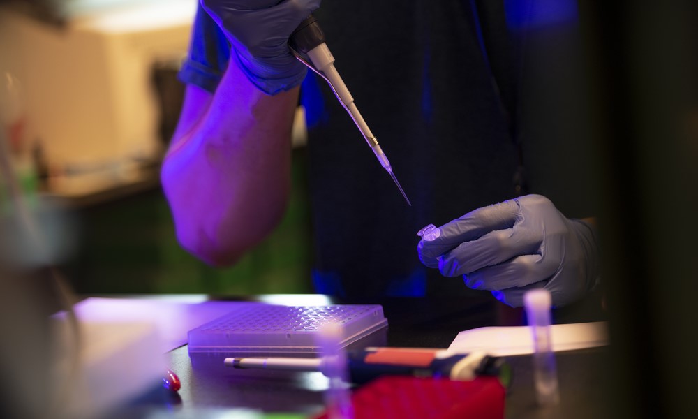
The primary role of the CID is genotyping patient tumors in an effort to direct molecularly targeted therapies, thereby enhancing the efficacy of targeted therapies as well as supporting prospective clinical trial designs. Patient analyses are performed in our CLIA-certified laboratory and results are generated within a timeframe that allows for timely treatment decisions. In addition, our laboratory pursues translational and clinical research studies to support the expansion of our genotyping profiles. To accomplish these goals, the lab utilizes many molecular and cellular techniques, including:
- Fluorescent in situ hybridization (FISH)
- Single gene molecular assays
- Next generation sequencing (NGS)
The combination of these approaches allows for the detection of somatic genetic aberrations, including gene copy number, point mutations and small insertions and deletions and RNA fusions.
Molecular Diagnostics & Mass General Pathology
In 2005, Mass General's Department of Pathology created the Molecular Diagnostics Laboratory under the direction of A. John Iafrate, MD, PhD. The test offerings started out small; the original two tests were 1P19Q fluorescent in situ hybridization (FISH) and microsatellite instability (MSI). The menu of tests has expanded significantly over time with the key addition of ALK FISH which functions to assist in lung cancer treatment.
In 2008, Dr. Iafrate created the Translational Research Lab (TRL) in collaboration with Mass General Cancer Center. The TRL’s mission was to design a multiplex test that would simultaneously test for the top cancer mutations for all cancer types. The resulting SNaPshot panel tests for 92 commonly mutated loci across 23 cancer genes.
In 2011, clinical molecular diagnostics was unified as the Center for Integrated Diagnostics (CID) at Mass General.
In 2013 the CID rolled out its first next generation sequencing (NGS) assay, AMP translocation, which looks for gene rearrangement of ALK, ROS, and RET. Building on past experience of targeting a specific number of genes in the original SNaPshot Panel and the NGS based technology of the AMP assay, we launched the first version of NGS-Snapshot in 2014.
The CID currently offer a variety of DNA and RNA-based NGS assays for solid tumors and hematologic malignancies, allowing comprehensive assessments for clinically actionable cancer genes.
Licensure & Accreditation
The Center for Integrated Diagnostics has CLIA (Clinical Laboratory Improvement Amendments) certification through CMS (Centers of Medicare and Medicaid Services) and is accredited by the Joint Commission. Please see the Pathology Department Regulatory Compliance website to request a copy of the CLIA certificate. The Center for Integrated Diagnostics is also a New York State CLEP (Clinical Laboratory Evaluation Program) licensed molecular laboratory for performing gene translocation assay, which can generate critical information for treating lung cancer patients. For more information about these assays, please contact the laboratory.
Leadership & Team
 Charlotte Wang, MD, PhD
Director (Interim), CID Lab, Massachusetts General Hospital
Assistant in Pathology, Massachusetts General Hospital
Instructor in Pathology, Harvard Medical School

Dora Dias-Santagata, PhD, FACMG
Associated Laboratory Scientist, Massachusetts General Hospital
Associate Professor of Pathology, Harvard Medical School
 Adam Fisch, MD, PhD
Assistant Pathologist, Massachusetts General Hospital
Assistant Professor of Pathology, Harvard Medical School
Researcher, Professor of Pathology, Massachusetts General Hospital
Austin L. Vickery, Jr. Professor of Pathology, Harvard Medical School
Assistant Professor of Pathology, Massachusetts General Hospital
Assistant Professor of Pathology, Harvard Medical School
Vice Chair for Pathology Informatics, Massachusetts General Hospital
Associate Clinical Director of Pathology Systems, Massachusetts General Hospital
Assistant Professor of Pathology, Harvard Medical School
Associate Director of Hematological Molecular Pathology, Massachusetts General Hospital
Associate Professor of Pathology, Harvard Medical School
Associate Professor of Pathology, Massachusetts General Hospital
Associate Professor of Pathology, Harvard Medical School
Assistant in Pathology, Massachusetts General Hospital
Instructor in Pathology, Harvard Medical School
 Julie Miller Batten, MLS (ASCP)
Technical Director, Center for Integrated Diagnostics
 Nick Jessop
Case Manager, Center for Integrated Diagnostics
Services
We provide a variety of cancer genotype testing and a limited number of non-cancer tests. Please see the Requisition Form below or contact the CID for more information.
Billing
Mass General Patients: Testing completed for Mass General patients will be billed directly to their insurance via their medical record number.
Non-Mass General Patients: Institutional invoice is the only billing option for non-Mass General patients. We cannot bill a patient’s insurance provider unless a patient has been seen by a Mass General clinician within 30 days of sample receipt. Please contact us for specific pricing information, as this is subject to change.
Get Started
In order for us to process a request for testing, all forms outlined in the Requisition Supplement (PDF) must be completed and submitted along with the specimen. Incomplete paperwork will be returned.
How to Ship: Any materials sent by either standard or express mail must be protected by using proper packaging. Glass slides should be enclosed in a protective slide box. Blood and bone marrow specimens should be in appropriate biohazard packaging and protected from extreme temperatures. All fluid specimens must be expedited to arrive within 5 days from date of draw. We suggest that all materials related to the case be shipped in the same container to ensure that they are received together. The mailing label should include a return address. Original H&E slides and blocks can be returned by our lab upon request. Unstained slides will be used for testing and will not be returned.
Support the Center for Integrated Diagnostics
The Massachusetts General Hospital Center for Integrated Diagnostics provides clinically actionable molecular diagnostics to accelerate the adoption of personalized medicine.


