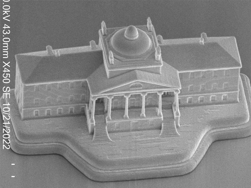
A familiar sight to MGHers is the Bulfinch Building, whether the actual granite edifice or depictions of it in historical photographs and logos of various eras.
Recent visitors to the laboratory of Guillermo (Gary) Tearney, MD, PhD, Department of Pathology, were treated to a new view of the building by peering through a scanning electron microscope.
A nano model of the Bulfinch Building.
The lab — part of the Wellman Center for Photomedicine — contacted staff of the hospital’s Paul S. Russell, MD Museum of Medical History and Innovation — no strangers to odd requests — to borrow a bread-loaf-size plaster model of the building. A three-dimensional scanning firm created a 3D computer-aided design model. Tearney Lab members then fed this information to the lab’s nanoscale printer to create a version that is 170 microns wide, about the width of a human hair.
Tearney chose to create this tiny Bulfinch — which possesses extraordinary detail, down to its five pillars and double staircase — as a compelling demonstration of the capabilities of their new printer, generously funded by donors.
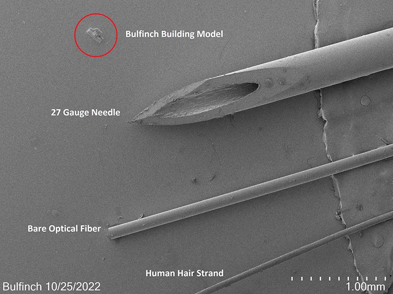
An exercise in scale: The Bulfinch Building — the real version of which dominated the neighborhood when it opened in 1821 — is dwarfed here by other items found in the lab.
The printer’s typical purpose is no less extraordinary: to create microscopic lenses that Tearney Lab members place inside medical imaging probes, which go on to be used inside patients in clinical trials. These devices allow clinicians to “fly through” the gastrointestinal tract, coronary arteries and lungs to see microscopic images inside living people and identify problems less invasively.
“One advantage of this nano 3D printer is we can now make tiny probes to image inside patients that are much smaller than can be made by hand,” says Tearney. “Also, we can make unique lens designs that cannot be made by any other manufacturing method, allowing the lenses to be used to grab images at unprecedented resolutions. We can now see, for example, microscopic cholesterol crystals in human coronary arteries that are harbingers of coronary artery disease.”
Through an international clinical observership initiative, Vadym Vus, MD, from the Ukraine and Andrew Liteplo, MD, from Mass General shared their focus of work and education in ultrasound imaging and diagnosis.
It takes a great deal of communication, coordination, flexibility and teamwork to manage the many patients who come in, and out, of the hospital each day. Within MGH Interventional Radiology alone, staff handle up to 70 cases per day – and this number is expected to steadily grow.