
Podcast5 Minute ReadNov | 1 | 2018
Extracellular Vesicles in Brain Cancer Blood Testing
- In this new study, Shannon Stott, PhD and Brian Nahed, MD, MSc of the Mass General Cancer Center describe a new technique that uses microfluidics and engineered nanosurfaces to capture EVs released from glioblastoma multiforme, the most common and aggressive form of brain cancer.
Extracellular Vesicles in Brain Cancer Blood Testing
Podcast
- Episode 47: Getting to know Shannon Stott, PhD, and Brian Nahed, MD, MSc
In this episode, Drs. Shannon Stott and Brian Nahed explain their backgrounds and share how they met and began working with each other. - Episode 48: A study on new techniques for capturing EVs released from glioblastoma with Shannon Stott, PhD, and Brian Nahed, MD, MSc
Drs. Shannon Stott and Brian Nahed describe a new technique that uses microfluidics and engineered nanosurfaces to capture EVs released from glioblastoma multiforme, the most common and aggressive form of brain cancer. - Episode 49: The impact and next steps for research led by Shannon Stott, PhD, and Brian Nahed, MD, MSc
Drs. Shannon Stott and Brian Nahed explain why their new technology is an important breakthrough for glioblastoma patients and share what’s next for their research.
Can a new screening for tiny cell particles help monitor the progress of brain cancers?
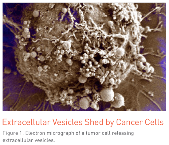 Extracellular vesicles (EVs) are a diverse group membranous structures that are shed from all cells of the body. They include exosomes, oncosomes, migrasomes and microvesicles, which play a role in cell-to-cell communication. Because cancer cells also shed these structures (Figure 1), and because they can be found in blood and other biological fluids, researchers are investigating whether they can capture cancer-related EVs as a noninvasive test for brain cancers at the time of diagnosis, through treatment and post-therapy.
Cancer cells can release up to 10,000 EVs per day. But since all cells release EVs, the question has remained how to isolate cancer-related EVs to retrieve the most information possible about a patient’s cancer status. In a paper published in Nature Communications in January 2018, Shannon Stott, PhD, Assistant Professor at the Center for Engineering in Medicine at Massachusetts General Hospital Cancer Center, and colleagues describe a new technique using microfluidics and engineered nanosurfaces to capture EVs released from glioblastoma multiforme (GBM), the most common and aggressive form of brain cancer. (1) “As a mechanical engineer, this is an area where I decided I could contribute to the field—by developing a technology that would pull out just the tumor-specific EVs,” says Dr. Stott, who is also a member of the BioMEMS Resource Center at Massachusetts General Hospital.
Shannon Stott, PhDThis technology is the next step for having a universal test for brain cancers, and we hope that it can better match patients to the most appropriate clinical trial.
Assistant Professor, Center for Engineering in Medicine, Massachusetts General Hospital Cancer Center; Assistant Professor of Medicine, Department of Medicine, Harvard Medical School
Microfluidic Chip Technology
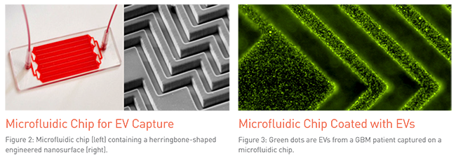
Since EVs are about 20,000 times smaller than a cell, the team needed to modify current techniques for moving bodily fluids through chip devices in order to capture accurate data at this scale. Stott’s technology works by manipulating interactions between blood particles and the nanoparticles on the device surface. “By using microfluidics, we are able to have very precise control of how these vesicles move through our technology,” says Dr. Stott, “ultimately enabling highly specific capture.”
By using a staggered herringbone pattern (Figure 2), the team was able to create chaos in the blood as it moved through the microfluidic chip. This resulting “chaotic flow” forms vortices in the fluid that increase the interactions between the vesicles and the engineered nanosurfaces of the device. In this proof-of- concept example, the EVHB-Chip device is lined with antibodies targeted against glioblastoma surface proteins. As the EVs from the glioblastoma cells flow through the chip they are captured by the antibodies on the surface of the device (Figure 3).
Proof of Concept in Glioblastoma
 In their study, the team, including Dr. Xandra Breakefield and Dr. David Ting, identified tumor-related EVs from 13 GBM patients. Furthermore, six of the patients possessed the epidermal growth factor receptor variant III (EGFRvIII) mutation, which is specific to GBM tumors, and the device was able to detect this specific mutation in five of the patients. The researchers were also able to perform RNA sequencing on these vesicles and identify hundreds of tumor specific genes. “We could look at these genes and map them to subtypes of tumors in glioblastoma as well as see markers of progression, angiogenesis and molecules that might give an indication of the aggressiveness of the tumor,” says Dr. Stott.
Dr. Stott and her colleague, Dr. Brian Nahed at Massachusetts General Hospital, have initiated a larger GBM study with this technology with the intention of following patients from the day of diagnosis throughout the course of their treatment—with the goal of trying to understand the clinical relevancy of the information from EVs. More than 50 patients have enrolled in this study with some receiving EV analysis up to 30 different time points during treatment. “It is early, but we are seeing dynamic responses in biomarkers throughout the patients’ treatments,” says Stott. “We are working to look at the correlation with their treatment responses.” Stott has focused on GBM initially because it is inherently difficult to source tissue from the brain, which is an obstacle to cancer screening. Early data suggests that EVs, in contrast to cancer cells or other, larger biomarkers of brain cancer, may be able to more readily pass the blood-brain barrier, which gives them the ability to convey important information in the blood about what’s going on there. “This technology is the next step for having a universal test for brain cancers,” says Dr. Stott, “and we hope that it can better match patients to the most appropriate clinical trial.” While this first application is in GBM, the team is also investigating the technology’s potential with other cancers, pancreatic vesicles and neurodegenerative diseases.
While this first application is in GBM, the team is also investigating the technology’s potential with other cancers, pancreatic vesicles and neurodegenerative diseases.
Reference
How to Listen to the Podcast
- Click on each episode below.
- Episode 47: Getting to know Shannon Stott, PhD, and Brian Nahed, MD, MSc
In this episode, Drs. Shannon Stott and Brian Nahed explain their backgrounds and share how they met and began working with each other. - Episode 48: A study on new techniques for capturing EVs released from glioblastoma with Shannon Stott, PhD, and Brian Nahed, MD, MSc
Drs. Shannon Stott and Brian Nahed describe a new technique that uses microfluidics and engineered nanosurfaces to capture EVs released from glioblastoma multiforme, the most common and aggressive form of brain cancer. - Episode 49: The impact and next steps for research led by Shannon Stott, PhD, and Brian Nahed, MD, MSc
Drs. Shannon Stott and Brian Nahed explain why their new technology is an important breakthrough for glioblastoma patients and share what’s next for their research. - Subscribe to the podcast series
- Click on your iPhone’s pre-loaded app called “Podcasts” (purple icon).
- Search for the series. Tap on the “search” icon, type in “Advances at Mass General Cancer Center,” and select it from the list of results.
- Subscribe. Once on the series page, tap on the “subscribe” button, and new episodes will be sent to your phone for free. You can adjust your settings to be alerted when a new episode is available.
Contributors
Shannon Stott, PhD
Assistant Professor, Center for Engineering in Medicine, Massachusetts General Hospital Cancer Center; Assistant Professor of Medicine, Department of Medicine, Harvard Medical School
Brian Nahed, MD, MSc
Neurosurgeon, Massachusetts General Hospital; Associate Director, Neurosurgery Residency Program, Massachusetts General Hospital; Associate Professor of Neurosurgery, Harvard Medical School
-
![]()
- Vice Chair of Education, MGB Department of Neurosurgery
- Neurosurgical Oncology / Brain Tumor Surgeon / MGH Brain Tumor Center
- Director, MGH Neurosurgery Residency Program

Krantz Family Center for Cancer Research
The scientific engine for discovery for the Mass General Brigham Cancer Institute.
We are home to more than 1,000 active clinical trials per year.
Ensuring that our patients have access to pioneering therapies.
Education & Training at the Cancer Institute
A world leader in cancer research & comprehensive cancer treatment, we are dedicated to educating the next generation of cancer specialists.