How TSC Affects the Eye
Tuberous sclerosis complex (TSC) can cause benign tumors in any major organ, including the eye. Approximately 50 percent of people with TSC exhibit at least one tumor, or lesion, inside the eye. Often these occur on the surface of the retina, the innermost layer responsible for receiving outside visual stimuli and transmitting that information to the brain.
Fortunately, most lesions of the retina and other TSC-related eye abnormalities usually do not grow and have little or no effect on the vision of people who have them. However, in many cases they serve an important role in the diagnosis of TSC, especially in the absence of many other signs of the disorder.
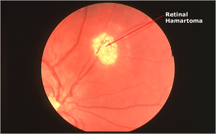
TSC-related tumors that form on the retina are called retinal hamartomas. Generally speaking, hamartomas are collections of abnormally shaped cells that multiply excessively to form benign tumors. In the eye, hamartomas are composed of abnormal neurons.
Other relatively minor eye manifestations of TSC include:
- Achromic patches, or spots on the retina that are lighter or darker in pigment than surrounding tissue
- Spots on the iris that are lighter in pigment than surrounding tissue—the eye equivalent of hypomelanotic macules (white patches) in the skin
- Light or depigmented eyelashes—the eye equivalent of light patches of hair, or poliosis, elsewhere on the body
- Angiofibromas that grow on the eyelids
- Small tumors that grow on the conjunctiva, the thin, transparent tissue that covers the outer surface of the eye
Diagnosis
In some people, TSC-related eye lesions represent the first signs of the disorder and are therefore important in diagnosis. Retinal achromic patches are found in 75 percent of people with TSC and are considered a minor feature of the disorder. Multiple retinal hamartomas are characteristic of TSC and are considered a major feature in the diagnostic criteria for TSC. It is important to note, however, that retinal hamartomas are clinically indistinguishable from eye lesions caused by another genetic disorder, neurofibromatosis. Therefore, doctors refrain from making a definite diagnosis on the basis of retinal lesions alone.
While some eye manifestations are near the surface and are easily observed, identifying retinal abnormalities typically requires examination by an ophthalmologist familiar with the signs of TSC. An ophthalmologic TSC evaluation usually involves pupil dilation and a technique called indirect ophthalmoscopy, which uses bright light and magnification to view the surface of the retina and identify any abnormalities. Retinal hamartomas have been seen in all ages of people with TSC, including infants. However, because the procedure can be uncomfortable, it may be difficult to perform on young children and cognitively impaired individuals.
Follow-up and Treatment
In rare cases, large retinal hamartomas can cause visual impairment. However, unlike many other TSC-related tumors, retinal hamartomas rarely increase significantly in size or number over time. As a result, people with TSC whose initial evaluations reveal only minor lesions seldom require repeated examinations.
In most cases, it is also unnecessary to treat retinal hamartomas and other TSC-related lesions of the eye. TSC specialists and ophthalmologists recommend only basic eye care for people with TSC who have no visual impairment or progressive hamartomas.
For cases in which vision is affected by large or progressive retinal hamartomas, treatments do exist. Ophthalmologists can eradicate hamartomas or reduce their size by eliminating the network of blood vessels that feed them using a technique called photocoagulation.
Next Steps
It is important to remember:
- TSC typically causes benign tumors in the eye
- These tumors, called retinal hamartomas, occur in approximately 75 percent of people diagnosed with TSC and may provide the first clinical signs of the disorder
- TSC may cause other eye abnormalities, such as light patches on the retina, spots on the iris, lightly pigmented eyelashes, angiofibromas on the eyelids and small tumors on the surface of the eye
- In most people with TSC, these eye manifestations cause no significant visual impairment
- What's more, retinal hamartomas and other tumors of the eye, unlike TSC-related tumors in other organs, tend not to progress over time
- Those who have been diagnosed with TSC or whose doctors suspect they have TSC should be examined by an ophthalmologist familiar with the disorder
- In cases where initial eye examinations show no sign of impairment, it is usually unnecessary to continue regular checkups
- In rare cases in which retinal hamartomas cause visual impairment, specialists may be able to reduce the size of tumors using a technique called photocoagulation