Press ReleaseJul | 27 | 2022
New Technology Developed by Mass General Researchers Creates a Multi-Color Molecular Movie
Key Takeaways
- Researchers have developed a new tool, scission-accelerated fluorophore exchange (SAFE) to see and differentiate between dozens of molecules on living cells
- Multiple color-coded images can be pieced together to produce a live, interactive movie of the molecules on cell surfaces
- The technology may be broadly useful for studying the functional dynamics between all types of cells without interfering with normal physiological processes
BOSTON – Many technologies have been developed with the goal of analyzing living systems in action at the molecular level. But they are often thwarted by the lack of visual details distinguishing the various molecules, or they destroy the cells in the process.
A new technology developed by researchers at the Massachusetts General Hospital sidesteps these problems. Called scission-accelerated fluorophore exchange (SAFE), the method uses various immunofluorescence tags attached to molecules on living cells, paired with uniquely fast yet exceptionally gentle chemical reactions to remove the tags.
 “The process allows us to see many different molecules at the same time in their natural state without destroying them,” said co-senior author Jonathan Carlson, MD, PhD, of the Center for Systems Biology at Mass General. “Layering all of the tagged molecules with different colors at the same time provides living detail, revealing cells and tissues moving, developing, and undergoing functional changes.”
Details of the technique are published in Nature Biotechnology.
The team’s goal was to develop a tool that lets them observe how cells interact and how tissues function. But when viewed through a microscope, it is difficult to discern between the molecules layering a cell’s surface because they are intrinsically nondescript in terms of any visual markers.
Though many forms of microscopy involve adding color to individual cells or molecules, current technologies lack the ability to produce rich arrays of molecules tagged in sequence in a sample that remains alive and intact.
“This is a technology that’s about seeing the flags that are flying on the surface of cells,” added Carlson, who developed the new chemical tools in collaboration with Hannes Mikula, now at the Institute of Applied Synthetic Chemistry in Vienna, and formerly a postdoctoral researcher in the laboratory of co-senior author Ralph Weissleder at Mass General.
“With SAFE, we can tag dozens of molecules, not just one or two, in the same system in a series of images collected over a short time, while that set of molecules and cells go on living,”
SAFE uses rapid bioorthogonal chemistry — chemical reactions that can be performed within living cells or systems that do not disrupt, interfere, or degrade the natural functions within them — to add and remove immunofluorescent signals from the surface of labeled cells.
The targets can be virtually any molecule on the surface of a cell, such as the key markers for distinguishing one cell type from another, or in classifying the diverse variety of immune cells.
The process uses nanomolar concentrations of two non-toxic reagents, a customized tetrazine and trans-cyclooctene pair, that act as chemical scissors that rapidly remove immunofluorescent tags during imaging.
“They are by far the brightest and the best tools when it comes to fast chemistry that's compatible with living systems,” explained Carlson, adding the chemicals are also gentle and require only small amounts to find each other, connect, and interact.
The team demonstrated that adding the fluorescent SAFE-labeled antibodies caused molecules to light up rapidly and fade as quickly as the fluorescent tags were added and removed. “The chemical scissors let us go from color to color or image to image quickly, all while the cells go on living,” said Carlson.
“The secret here is not that we can erase colors, it's that we can do so with exceptional speed, gentleness, and efficiency in a manner that the tissue doesn't even notice it's happening.”
The team showed that more than 99% of the color signals were removed in less than 30 seconds. SAFE overcomes limitations of existing technologies that use harsh chemicals that destroy the color, and the molecule or cell in the process.
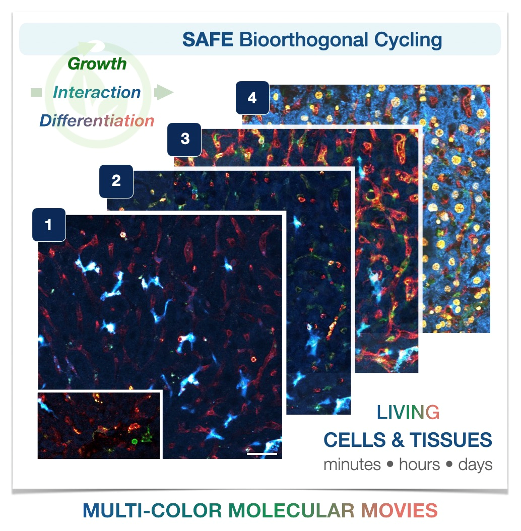
In this paper, the researchers used SAFE to create a movie-like continuous stream of multi-color images showing the migration of immune cells within living mouse liver tissue.
They also created multiplexed 14-color images of living mouse bone marrow and tracked the maturation of bone marrow progenitor cells as they differentiated into neutrophils over a six-day period, fully exchanging their cell surface ‘flags’ in the process .
“We anticipate that SAFE will provide a broadly useful new ability to visualize complex living systems across space and time,” said Carlson. “We envision it will provide significant new insights into both healthy physiology and the inner workings of diseases, particularly cancer, where the dynamics of tumor cells and the immune system are an area of great interest.”
Additional study authors include first author Jina Ko (now at the University of Pennsylvania), Martin Wilkovitsch, Juhyun Oh, Rainer H Kohler, Evangelia Bolli, Mikael J Pittet, Claudio Vinegoni, and David B Sykes.
This work was supported, in part, by grants from the CSB development fund and the National Institutes of Health.
About the Massachusetts General Hospital
Massachusetts General Hospital, founded in 1811, is the original and largest teaching hospital of Harvard Medical School. The Mass General Research Institute conducts the largest hospital-based research program in the nation, with annual research operations of more than $1 billion and comprises more than 9,500 researchers working across more than 30 institutes, centers and departments. In August 2021, Mass General was named #4 in the US News & World Report list of "America’s Best Hospitals."