Multiple Sclerosis Imaging Laboratory


MS Imaging Laboratory
Eric Klawiter, MD, MSc
Charlestown Navy Yard, Building 149
149 13th Street
Charlestown,
MA
02129
Phone: 617-726-7531
Email: klawiter@nmr.mgh.harvard.edu
Explore This Lab
Overview
The Multiple Sclerosis (MS) Imaging Lab, directed by Eric Klawiter, MD, MSc, focuses on MS clinical research as well as the application and development of new imaging methods to better understand, diagnose and treat MS.
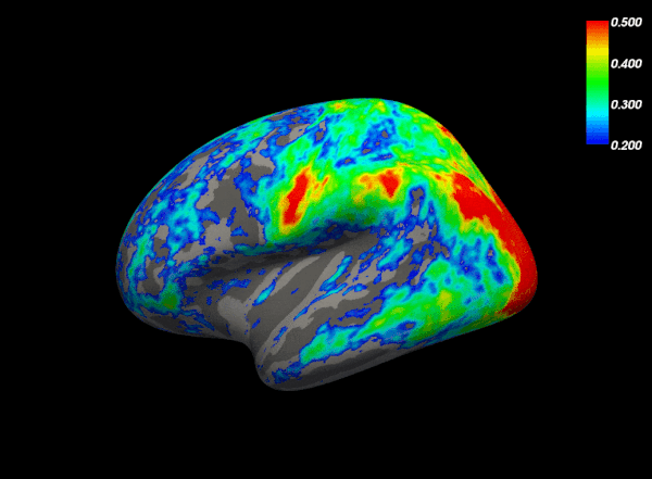
Through the use of novel imaging techniques, we examine changes in structural and functional brain networks in MS in relation to clinical outcomes such as cognition. The overall goal is to integrate novel imaging techniques into therapeutic clinical trials, which our lab is also involved with.
The goals of our research are to:
- Understand the pathology that gives rise to symptoms and cognitive impairment associated with MS
- Develop biomarkers for early diagnosis of MS
- Develop more effective treatments for MS
Our current imaging dataset consists of data from the 3T Connectom scanner and an ultra-high field 7T scanner on patients with MS including:
- Longitudinal structural magnetic resonance imaging (MRI)
- Longitudinal resting state functional MRI
- Diffusion tensor imaging (DTI)
- Advanced diffusion imaging (i.e. Q-ball and axon diameter modeling)
- Neurocognitive testing
- Optical coherence tomography (OCT)
- Clinical data
We also plan, implement and write customized code to streamline data processing and analysis.
Research Projects

Multi-modal Neuroimaging
The MS Imaging Laboratory is involved in various neuroimaging studies that implement established and novel techniques to examine structural and functional connectivity in relationship to cognitive dysfunction in MS.
Spinal Cord Imaging
We are exploring novel spinal cord imaging techniques in MS and related diseases such as neuromyelitis optica (NMO).
Selected Publications
Structural disconnection is responsible for increased functional connectivity in multiple sclerosis.
Patel KR, Tobyne S, Porter D, Bireley JD, Smith V, Klawiter E. Brain Struct Funct. 2018 Jun;223(5):2519-2526. doi: 10.1007/s00429-018-1619-z.
Corpus callosum axon diameter relates to cognitive impairment in multiple sclerosis.
Huang SY, Fan Q, Machado N, Eloyan A, Bireley JD, Russo AW, Tobyne SM, Patel KR, Brewer K, Rapaport SF, Nummenmaa A, Witzel T, Sherman JC, Wald LL, Klawiter EC. Ann Clin Transl Neurol. 2019 Mar 30;6(5):882-892. doi: 10.1002/acn3.760.
A surface-based technique for mapping homotopic interhemispheric connectivity: Development, characterization, and clinical application.
Tobyne SM, Boratyn D, Johnson JA, Greve DN, Mainero C, Klawiter EC. Hum Brain Mapp. 2016 Aug;37(8):2849-68. doi: 10.1002/hbm.23214.
Cognitive impairment and the regional distribution of cerebellar lesions in multiple sclerosis.
Tobyne SM, Ochoa WB, Bireley JD, Smith VM, Geurts JJ, Schmahmann JD, Klawiter EC. Mult Scler. 2018 Nov;24(13):1687-1695. doi: 10.1177/1352458517730132.
Our Research Team
-
![]()
- Director, Multiple Sclerosis and Neuromyelitis Optica Unit

Support Our Work
Philanthropic support for the Mass General Department of Neurology is critical to patient care, research and education. Please consider a gift to support neurology research and clinical care today.